
Quick reference
| Question | What to expect |
|---|---|
| Which projections are confirmed? | PA, lateral, and oblique elbow views. |
| Can users try other projections? | Yes. These are confirmed examples. Other projections may be possible when the simulated setup supports them. |
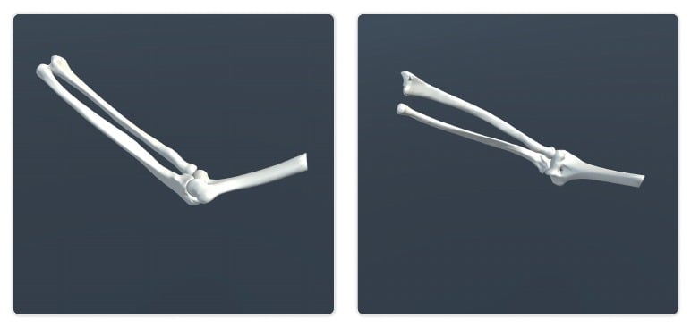
| What anatomy is included? | Humerus, radius, and ulna. |
| What setup is used? | Free-plating workflow. |
| What movement is included? | Shoulder internal and external rotation, elbow flexion and extension, and patient anteroposterior and mediolateral adjustment. |
| What bone variations exist? | Healthy bone version, distal humerus fracture, radial head fracture, and ulnar olecranon fracture. |
| Known limitation | Forearm supination and pronation are not included. |
Visual examples
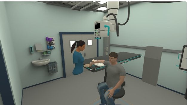
- Simulator setup
- Bone model
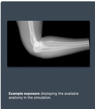
- Example exposure
Imaging capabilities
| Capability | Details |
|---|---|
| Radiographic tube | Full range of motion, including vertical, horizontal, tilt, and rotation. The cassette can be rotated, and collimation size is adjustable. |
| Detector methods | The detector setup uses free plating. |
| Simulated exposures | Confirmed support for PA, lateral, and oblique elbow views. Additional views may be possible depending on patient range of motion and available bones. |
Anatomy and movement
| Area | Details |
|---|---|
| Included anatomy | Humerus, radius, and ulna. |
| Joint mobility | Shoulder internal and external rotation. Elbow flexion and extension. Patient anteroposterior and mediolateral adjustment. |
Bone variations
- Healthy bone version
- Distal humerus fracture
- Radial head fracture
- Ulnar olecranon fracture