Quick reference

Projection expectations

The projections listed on this page are confirmed examples for this simulation module. They do not define the full limit of what can be attempted. The simulator creates an image from the selected anatomy, patient position, joint movement, detector setup, tube position, collimation, and exposure settings, so other projections may be possible when the setup supports them.

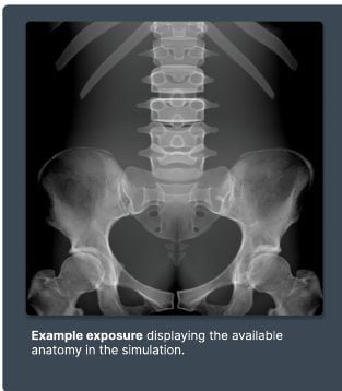
Visual examples

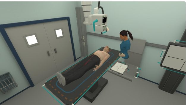
Simulator setup preview

Simulator setup and positioning environment
Imaging capabilities

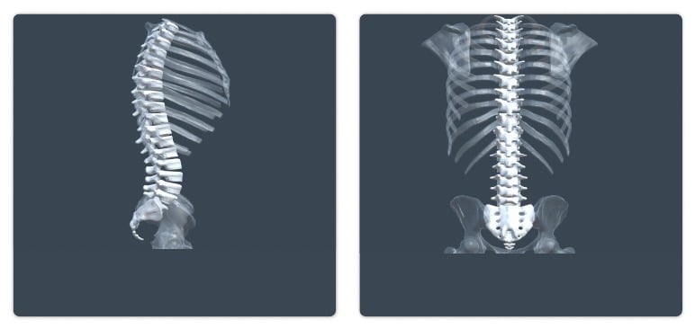
Anatomy and movement

Bone variations

  • Healthy bone version

Known limitations

The L-spine simulation is limited to recumbent patient positioning. This restricts projections that require prone or erect positioning. The vertebrae also have a low presence of trabecular bone structures, which may limit radiographic realism when evaluating bone quality and internal architecture.