
Quick reference
| Question | What to expect |
|---|---|
| Which projections are confirmed? | PA, lateral, and oblique elbow views. |
| Can users try other projections? | Yes. These are confirmed examples. Other projections may be possible when the simulated setup supports them. |
| What anatomy is included? | Humerus, radius, and ulna. |
| What setup is used? | Free-plating detector workflow. |
| What movement is included? | Shoulder internal/external rotation, elbow flexion/extension, and patient anteroposterior and mediolateral adjustment. |
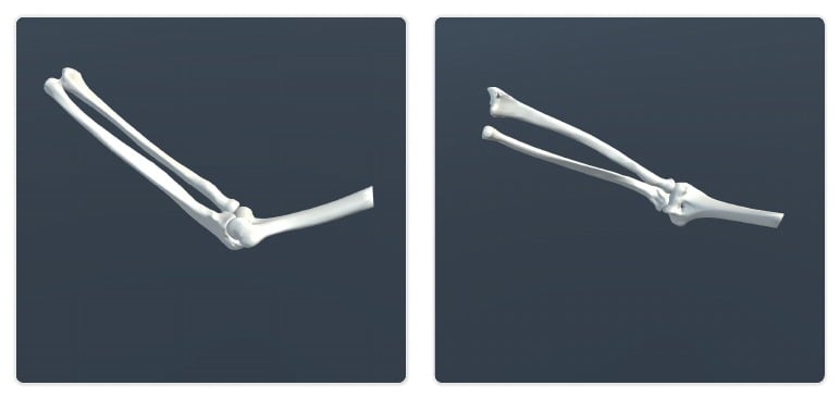
| What bone variations exist? | Healthy bone version, distal humerus fracture, radial head fracture, and ulnar olecranon fracture. |
| Known limitation | Forearm supination and pronation are not included. |
Visual examples
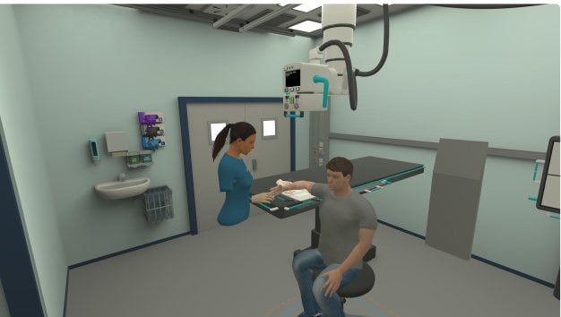
- Simulator setup
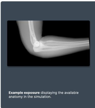
- Example exposure

Imaging capabilities
| Capability | Details |
|---|---|
| Radiographic tube | Full range of motion, including vertical, horizontal, tilt, and rotation. The cassette can be rotated, and collimation size is adjustable. |
| Detector methods | The detector setup uses free plating. |
| Confirmed projections | Confirmed examples include PA, lateral, and oblique elbow views. They do not define the full limit of what can be attempted. Other projections may be possible when the simulated setup supports them. |
Anatomy and movement
| Area | Details |
|---|---|
| Included anatomy | Humerus, radius, and ulna. |
| Joint mobility | Shoulder internal and external rotation. Elbow flexion and extension. Patient anteroposterior and mediolateral adjustment. |
Bone variations
- Healthy bone version
- Distal humerus fracture
- Radial head fracture
- Ulnar olecranon fracture